Vol.
30 No. 2
March-April 2008
The Human Kinome—Invitrogen Corp. Poster Caption
Phosphorylation by protein kinases represents a central theme within signal transduction. Since the description of reversible protein phosphorylation influencing protein activity by Fischer and Krebs in 1955, protein kinases have been shown to play central roles in the regulation of most critical cellular processes. Given the importance of the coordinated action of these enzymes, it is not surprising that misregulation of protein kinases is a cause or consequence of many diseases.
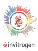
This circular diagram represents an artistic interpretation of the phylogenetic relationships between all members of the human kinase family. Our bioinformatics analysis concluded that the superfamily comprises 517 distinct protein kinases (including the 14 enzymes containing two kinase domains; 531 catalytic domains in all). This is slightly different from the description provided by Manning and colleagues (2002, Science 298:1912) in which 518 kinases and 13 secondary kinase domains were defined.
The kinases are organized into nine major groups (TK, TKL, STE, CK1, AGC, CMGC, CAMK, Other, and Atypical). The nine major groups are based upon the similarity between the amino acid sequences of the enzyme catalytic domains, with two exceptions. The Other group contains a highly divergent set of kinases that have significant similarity to the typical protein kinase domain; the Atypical group has insignificant sequence similarity to the typical protein kinase domain. The groups are further classified using multiple-sequence alignment programming with phylogenetic analysis to create the representative dendrograms.
These nine major groups are labeled and colored distinctly on the diagram. Each kinase protein is designated on the diagram as a single bar. The length of the bar is proportional to the number of branch points leading to the kinase. Alternating shades depict clusters within each group. Each kinase protein is named according to the HUGO Gene Nomenclature Committee (HGNC) naming convention, followed by the name used by Manning et al. if different.
The three-dimensional structure in the middle of the diagram is a model based on cAMP-dependent protein kinase. The red ribbon represents the backbone structure of the kinase. The yellow ribbon represents a 20–amino acid peptide inhibitor (PKI 5–24) bound to the kinase. The green model is AMP-PNP (an ATP analog) bound to the kinase active site. The two blue dots represent the two active-site Mg2+ ions necessary for the kinase activity.
©2007 Invitrogen Corporation.
www.invitrogen.com/kinasebiology
Page last modified19 March 2008.
Copyright © 2003-2008 International Union of Pure and Applied Chemistry.
Questions regarding the website, please contact [email protected] |